

Ultrasound scans are a crucial part of prenatal care, offering more than just a peek at your baby. These scans help monitor the baby's development and identify potential issues early on. It can help in detecting conditions like heart defects, spina bifida, and other anomalies that may affect the baby’s development. We spoke to our expert Dr Rama Krishnan, Radiologist - Neuberg Diagnostics, Chennai, who explained how these scans work, what they can reveal, and why early detection is so important for ensuring the best outcomes for both you and your baby.
Table of Content:-
What is Ultrasound?

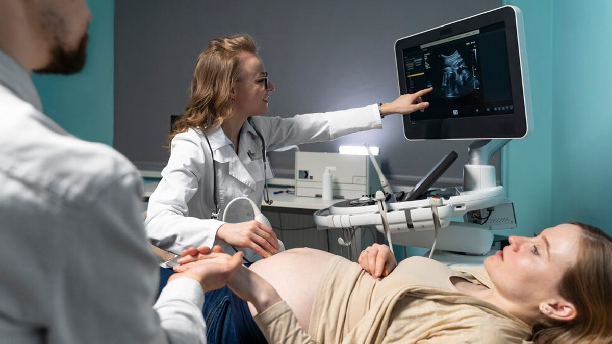
Ultrasound is a medical imaging technique that uses high-frequency sound waves (>20,000 Hz) to create images of structures within the body. "During pregnancy, these sound waves visualise the foetus in real-time providing crucial information about the baby’s growth, organ development, and overall well-being. Also, ultrasound is non-ionising and safe for both mother and foetus," said Dr Krishnan.
According to a 2023 study, prenatal ultrasound offers many practical advantages, such as allowing parents to prepare, plan for delivery, and ensure the best paediatric care for their baby. The first ultrasound is typically done between the 11th and 13th weeks of pregnancy, the second between the 20th and 21st weeks, and the third between 34-36 weeks for women with higher-risk pregnancies.
Also Read: Doctor Explains The Role of Ultrasound Scans in Pregnancy
Early Detection of Foetal Anomalies

“The Nuchal Translucency (NT) Scan, performed using ultrasound between 11-14 weeks of pregnancy, is an important prenatal screening test used to assess the risk of certain chromosomal abnormalities and structural anomalies in the developing foetus,” said Krishnan.
The NT scan measures the clear space in the tissue at the back of the baby's neck, known as nuchal translucency. An increased thickness in this area may be associated with a higher risk of chromosomal abnormalities (Down Syndrome, trisomy 18, etc) and congenital heart defects.
Common Anomalies Detected By Ultrasound
The dedicated foetal anomaly scan is typically conducted during the second trimester around 18-22 weeks. It allows doctors to assess the baby’s organs, limbs, and other key structures to identify potential abnormalities. These include:

- Neural Tube Defects: Anomalies, such as spina bifida, where the neural tube does not close completely.
- Heart Defects: Congenital heart defects, including septal defects or abnormalities in heart valves, can be diagnosed through a detailed foetal echocardiogram.
- Facial Anomalies: Cleft lip/palate and abnormally small jaw.
- Limb Malformations: Limb-related deformities like clubfoot or missing limbs.
- Gastrointestinal Anomalies: Conditions, such as gastroschisis or omphalocele, where abdominal organs develop outside the body.
- Genitourinary Anomalies: Conditions, such as renal agenesis (absent kidney), and hydronephrosis.
- Skeletal Dysplasia: Inadequate bone formation/development like achondroplasia.
- Multiple Pregnancy Complications: Twin-to-Twin Transfusion Syndrome (TTTS), a condition in identical twin pregnancies where blood flow between twins is uneven, leading to growth disparities
Also Read: Understanding Congenital Anomalies: Symptoms, Causes, And Prevention Tips For Birth Defects
Importance of Ultrasound in High-Risk Pregnancies
For women with high-risk pregnancies, such as those with a history of birth defects or other medical conditions, ultrasound is crucial in close monitoring. “Specialised ultrasounds like the level II ultrasound, which offers more detailed images, are often used to provide a thorough examination of the foetus. In some cases, conditions can be treated in utero, while others may require specialised care immediately after delivery. Early awareness can improve outcomes for both mother and baby,” said Dr Krishnan.
Bottomline
Dr Krishnan concluded, “Ultrasound plays an essential role in detecting foetal anomalies, offering early and accurate diagnosis of various conditions. This technology not only helps in guiding pregnancy management but also provides parents with crucial information to prepare for any potential challenges post-birth. With advancements in ultrasound technology, its effectiveness in foetal anomaly detection continues to improve, ensuring better outcomes for both mother and child.”
[Disclaimer: This article contains information provided by an expert and is for informational purposes only. Hence, we advise you to consult your own professional if you are dealing with any health issues to avoid complications.]
Also watch this video
How we keep this article up to date:
We work with experts and keep a close eye on the latest in health and wellness. Whenever there is a new research or helpful information, we update our articles with accurate and useful advice.
Current Version