

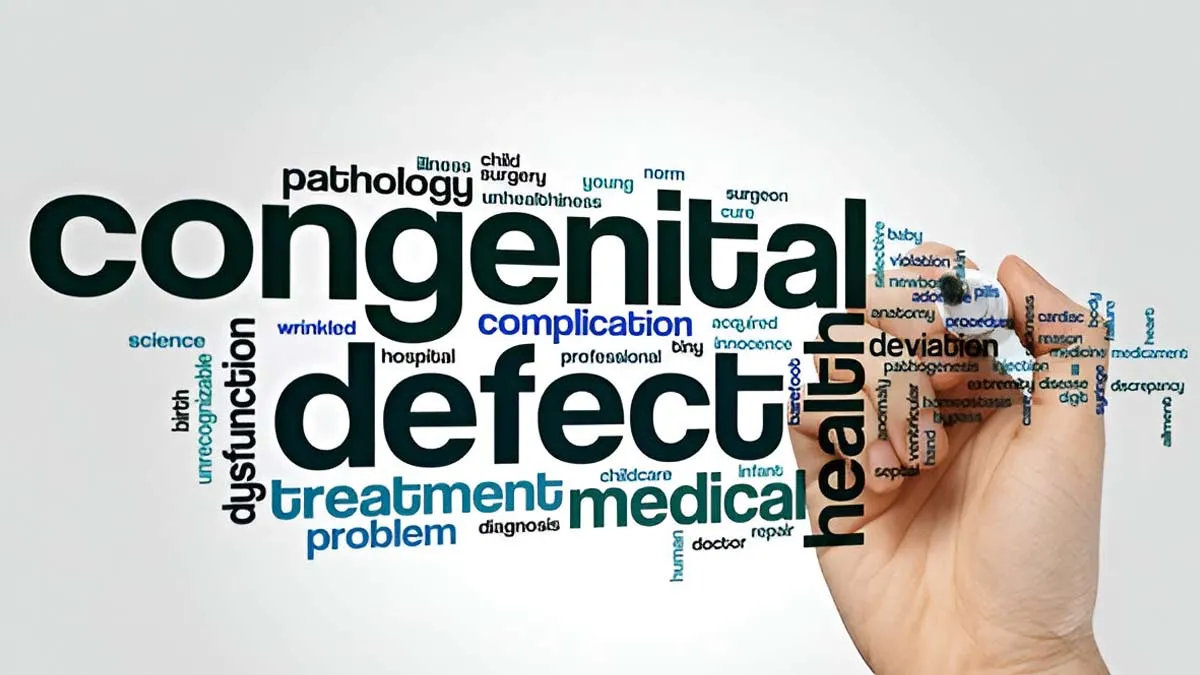
Congenital Heart Defects (CHD) are among the most common birth anomalies worldwide, affecting roughly one in every 100 newborns. Some defects are minor and require little intervention; others are life-threatening in the first hours or days of life. The difference between a safe outcome and a crisis often depends on timing — rapid recognition, accurate diagnosis, and timely referral can alter the course of an infant’s life.
We spoke to Dr Prashant Prakashrao Patil, Senior Consultant, Interventional Pediatric Cardiologist, CARE Hospitals, Banjara Hills, Hyderabad, who explained how early detection can save infants with cardiac defects.
Why Early Detection Matters?

"Several cardiac lesions present subtly at birth, with normal colour or breathing initially, and then deteriorate as transitional circulation changes occur. Early detection reduces the likelihood of emergency transfers, decreases preoperative instability, and improves readiness for neonatal interventions," said Dr Patil.
According to a 2016 study, globally, the prevalence of congenital heart disease (CHD) is estimated at 8 to 10 per 1,000 live births, though it varies significantly across different regions. Heart defects account for 28% of all major congenital anomalies.
When a defect is recognised before clinical collapse, the care team can optimise oxygenation, avoid inappropriate therapy, and plan safe transport to specialised centres — steps that reduce short-term complications and set the stage for better long-term outcomes.
Also Read: Ventricular Septal Defect In Newborns: What It Is And How To Treat It?
Prenatal Diagnosis: Spotting Trouble Before Birth
Foetal echocardiography and detailed obstetric ultrasound have improved the ability to detect structural heart problems during pregnancy. Prenatal diagnosis creates a window for planning:
- Delivery in a tertiary centre with paediatric cardiology on site
- Anticipatory counselling for parents, and
- Immediate postnatal management protocols
“In selected high-risk lesions, antenatal recognition shortens the time to definitive care and reduces the need for emergency procedures in unstable neonates. The sensitivity of foetal imaging varies by lesion and operator experience, but its growing role in perinatal planning is now central to modern paediatric cardiac services,” added Dr Patil.
Newborn Screening: Pulse Oximetry and the Bedside Exam
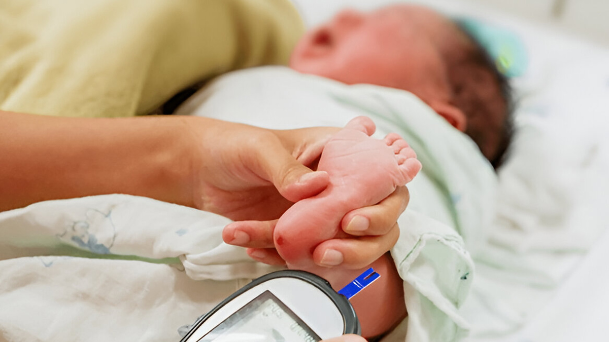
Since its inclusion on recommended newborn screening panels, pulse oximetry screening for critical congenital heart disease (CCHD) has become a routine, low-cost tool. Measuring oxygen saturation in the right hand and either foot after 24 hours of life helps identify cyanotic lesions that might otherwise go unnoticed.
Pulse oximetry does not replace clinical examination or imaging, but when combined with careful newborn assessment and prenatal data, it substantially increases the detection of life-threatening cardiac lesions before discharge. National clinical bodies continue to refine algorithms to reduce false positives while maximising sensitivity.
Imaging and Minimally Invasive Options Have Sharpened Diagnosis and Therapy
Echocardiography remains the diagnostic cornerstone, but advances in neonatal imaging, such as higher-resolution probes, three-dimensional reconstructions, and point-of-care machines, allow more precise anatomical and functional assessment at the cot-side.
“Where anatomy permits, catheter-based interventions now treat conditions that once required open surgery. Transcatheter closure of the ductus arteriosus in selected preterm infants and device closure of some septal defects are examples of minimally invasive approaches that shorten hospital stay and reduce wound-related complications. These techniques demand specialised expertise and appropriate equipment, but they expand the therapeutic window for fragile neonates,” explained Dr Patil.
Also Read: Can You Reduce The Risk Of Birth Defects? Common Questions Answered By Expert
Surgical Advances and Intensive Neonatal Support
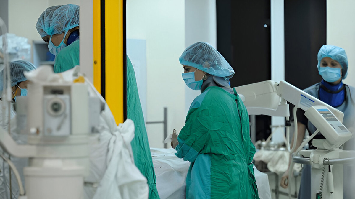
When surgery is required, neonatal cardiac surgery has benefitted from refinements in bypass technology, myocardial protection, and perioperative critical care. Hybrid strategies, surgical and catheter techniques, are increasingly used for complex lesions, offering staged solutions that reduce immediate physiologic stress.
Extracorporeal Membrane Oxygenation (ECMO) and advanced neonatal intensive care provide life-saving support for infants who develop cardiorespiratory failure pending repair or recovery from critical conditions. These advances have shifted outcomes: selected cohorts with previously grim prognoses now survive to discharge and enter long-term follow-up programmes.
What Clinicians and Families Should Expect
A coordinated pathway is crucial and should include:
- Prenatal screening
- Routine pulse oximetry
- Prompt echocardiography for abnormal findings
- Timely referral to paediatric cardiology or cardiothoracic surgery
Families should receive:
- Clear counselling about likely trajectories
- Information about possible interventions
- Explanation of the structure of follow-up care
- Empathetic support
For clinicians in non-specialist settings, the priority is stabilisation and early transfer to centres with neonatal cardiac and intensive care capacity rather than attempting definitive procedures without appropriate resources.
Bottomline
Dr Patil concluded, “Early detection transforms congenital cardiac care. When a cardiac lesion is recognised before clinical collapse, teams can plan delivery, anticipate needs, and match the infant to the right intervention at the right time. The combined progress in prenatal imaging, newborn screening, catheter techniques, surgical care, and neonatal support is shortening emergency response times and improving survival and quality of life for affected infants. Continued investment in training, screening implementation, and regional pathways will ensure that more babies born with a brave heart have the best possible start.”
Also watch this video
How we keep this article up to date:
We work with experts and keep a close eye on the latest in health and wellness. Whenever there is a new research or helpful information, we update our articles with accurate and useful advice.
Current Version