

For the diagnosis of secondary bone cancer, initially a blood test is done to check the calcium level in the blood. Hypercalcaemia, high levels of calcium in the blood, is associated with malignancy such as multiple myeloma, breast and lung cancer, which spread to the bone as secondary bone cancer.
 X-rays of the chest to check for cancer in the lungs and bones of the individual are done next to check for any abnormality, but may this may not show up clearly seen in x-ray images.
The third series of tests for secondary bone cancer diagnosis is done by injecting a small amount of a mild radioactive substance into the patient. This is called ‘bone scanning’. After about 2-3 hours after the injection, the radioactive substance gets absorbed by the bones affected by secondary bone cancer and can be seen clearly in x-ray images as hot spots.
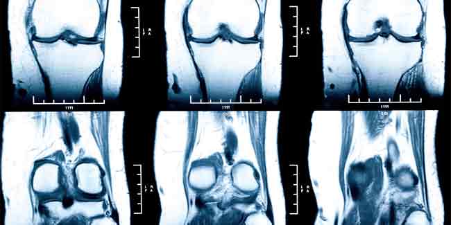
A CT (Computerised Tomography)scan and MRI (Magnetic Resonance Imaging) that show3D images and can help doctors identify if the patient is suffering from secondary bone cancer and pinpoint the location of the cancer to be able to direct radiotherapy at that location.
A newer method of diagnosing secondary bone cancer is the PET scan (Positron Emission Tomography) in which a low dose of radioactive glucose is given to the patient that can identify the cell activity in different parts of the body and help identify the bones affected by secondary bone cancer.
Very often even after these tests doctors may not be still sure of the cause of the abnormality in the bone that caused secondary bone cancer and ask for a bone biopsy, to study the cells taken from the affected area of the bone. This can be done in one of the following ways:
- Needle biopsy is a test in which a sample of the bone is taken to check for the nature of cancer cells by passing a needle through the skin into the bone and extracting a small piece of the bone for microscopic examination. A local anaesthetic may be needed to numb the area and help the patient relax.
- Open biopsy is done under general anaesthesia as a small piece of bone has to be extracted for examination. Bones are extremely hard and the removed piece has to be softened to examine it under a microscope. This takes several days and the patient has to wait for about two weeks before the result is out.
Cancer cells in the bones of the secondary bone cancer are the same as those of the organ from where the primary cancer has metastatised. It may sometimes happen that the secondary bone cancer has been found before the primary cancer is diagnosed. In such a case doctors will ask for the following tests:
- A mammogram to look for primary cancer in the breast
- A chest x-ray to see if there is lung cancer
- A CT or ultrasound scan to check for kidney cancer
- Prostrate ultrasound and a blood sample to see if prostate cancer is present.
All these tests may take a few weeks to accomplish, but it is important to make an accurate diagnosis.
In conclusion diagnosis of secondary bone cancer is a long process but essential to doctors to chart a course of curative action.
How we keep this article up to date:
We work with experts and keep a close eye on the latest in health and wellness. Whenever there is a new research or helpful information, we update our articles with accurate and useful advice.
Current Version